What Potassium Does to the ECG, Interactive Simulation
Of all the electrolytes, potassium is the one I would least want to get wrong. Slide it from 1.5 to 9.5 mmol/L and watch what the heart does.

Learning by doing, safely#
Interactive simulations are changing medical education by replacing passive learning with safe, hands-on practice. The learner does not read about a hyperkalemic ECG. The learner makes one: move a control, and the model answers at once. Cause and effect sit on the same screen. TriageCode MD is pleased to present a computer-driven interactive simulation of the electrocardiogram (ECG) in potassium disorders. One slider runs from 1.5 to 9.5 mmol/L, and the tracing rebuilds itself as the number changes. It is a teaching schematic, not a patient record, and it says so on the screen.
Try the interactive simulator#
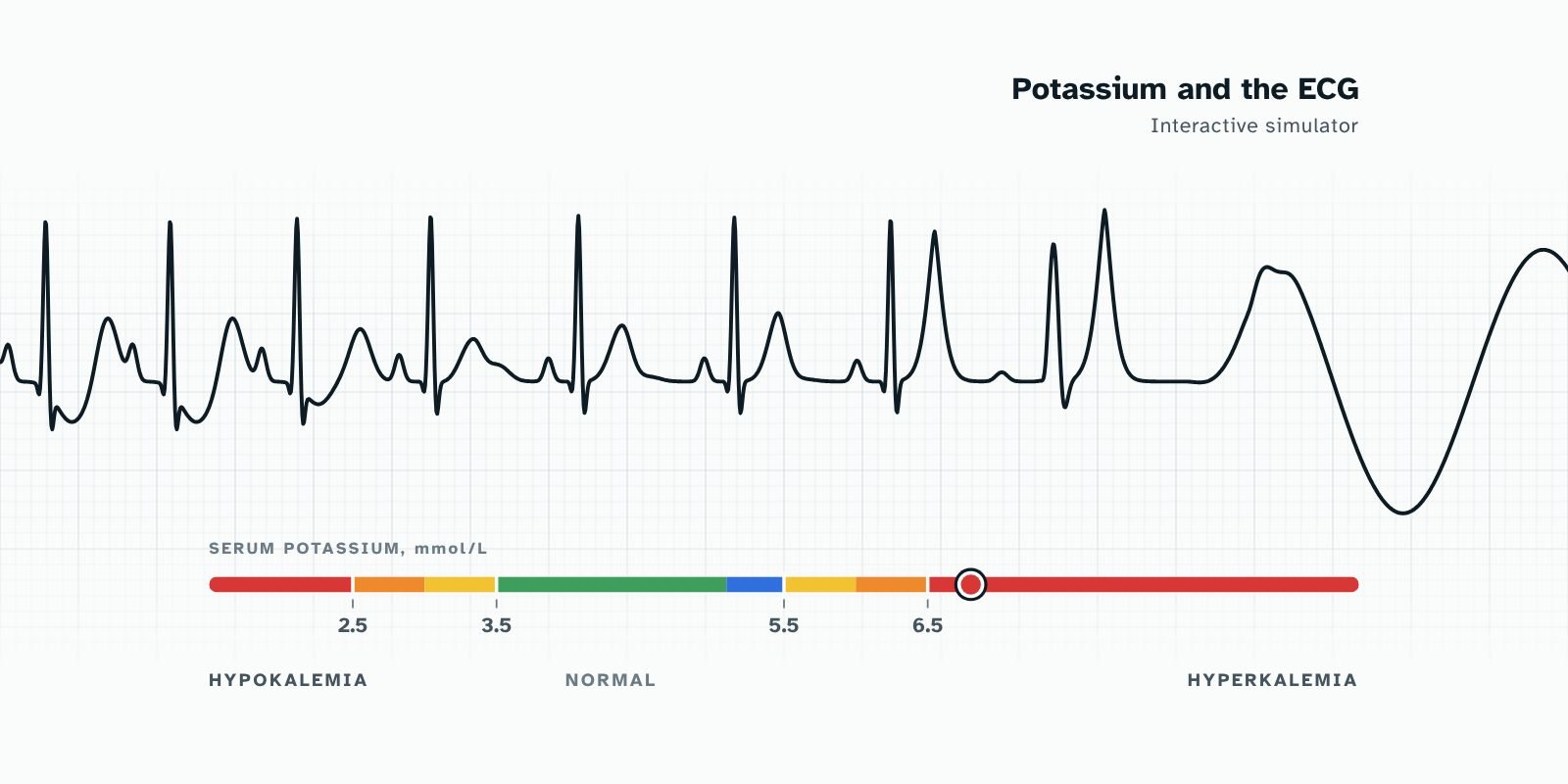
Slide the bar, press the buttons and try to break it. The ECG moves with the potassium, and the two boxes, ECG findings and signs and symptoms, change colour with severity: red is life-threatening, orange urgent, yellow caution, green normal, blue borderline, so recheck it. Each colour also has a word and an icon, so you never depend on colour alone. Start at 4.2, normal, and note the PR and QRS numbers. Slide up from 5.0: between 5.5 and 6.0 the T wave turns tall and narrow. Stop at 6.0, where the UK guideline asks inpatients for an urgent 12-lead ECG 1. At 7.5 the P wave is almost gone, the PR is long and the QRS number climbs. At 9.0 comes the sine wave: the heart is about to stop. Now go the other way. At 3.0 the ECG may look almost normal, which is the trap. Slide down through 2.7 and watch the T wave flatten, the ST segment sag and a U wave grow; below 2.0 the T and U waves fuse. Finally, press "Label waves", then "Test me": the potassium is hidden and you pick the range from the ECG.
Interactive example: Potassium and the ECG: interactive simulator. It needs JavaScript to run. Open the article to try it.
What the simulator shows#
A textbook gives you stills: one strip at 6.5, another at 8.0. A heart does not behave in stills. In the simulator each wave responds to potassium in its own way, and you watch it happen.
Raise the potassium and the T wave grows tall and narrow. The P wave flattens and fades, the PR interval stretches, and the QRS widens until it merges with the T wave into a sine wave. Lower it and the T wave flattens, the ST segment sags and a U wave appears. Go lower still and the T and U waves fuse, so the QT looks long when it is really a QU interval.
Readouts for heart rate, PR, QRS and corrected QT change under the strip as you move, so shape and number are learned together. A "Label waves" button names the P, QRS, T and U on the tracing.
The order matters most. In hyperkalemia the T wave changes first, then the P wave and PR interval, then the QRS. A table can state that sequence. A slider lets you see it happen.
Why simulation helps#
Physicians read ECGs imperfectly. A meta-analysis of 78 studies found that accuracy was low without training and varied widely 2. Simulation is one way to supply that training. Across 609 studies and 35,226 learners, technology-enhanced simulation was associated with large effects on knowledge, skills and behaviours compared with no intervention, and moderate effects on patient-related outcomes 3. A smaller review of 14 studies found simulation with deliberate practice beat traditional clinical education for skill acquisition 4.
What does interactivity add?
| What it adds | How it works in the simulator |
|---|---|
| Immediate feedback | The tracing answers the moment you move. |
| Repetition without risk | A wrong guess costs nothing, and you can run the same range 20 times. |
| Control | The learner sets the pace and chooses what to explore. |
| Retrieval | In "Test me" mode the potassium is hidden. You read the ECG, choose the range and learn at once whether you were right. That asks you to pull the pattern from memory rather than look it up. |
Who it is for#
Clinicians can rehearse the pattern before the next patient with a high potassium and an odd-looking ECG. Academics get a demonstration that is the same for every class, needs no equipment or login, stores nothing about the learner and runs on a phone. Researchers get a parametric model: each wave is a handful of numbers, so the assumptions are open to inspection and challenge, and the model could supply a standard set of tracings for studies of ECG recognition.
Key point
Simulation has not been shown to beat a good lecture for ECG teaching. In a randomized trial, a computer simulator and traditional teaching gave similar test scores immediately and at three months 5. I make no claim that this simulator raises scores, and real ECGs are messier than the schematic. Its case is practice, immediacy and reach, added to the teaching you already do. To what extent do interactive simulations enhance the diagnostic accuracy of electrocardiogram interpretation?
References
- UK Kidney Association. Clinical practice guidelines: treatment of acute hyperkalaemia in adults. 2023 update. ↩︎
- Cook DA, Oh SY, Pusic MV. Accuracy of physicians' electrocardiogram interpretations: a systematic review and meta-analysis. JAMA Intern Med. 2020;180(11):1461-71. doi:10.1001/jamainternmed.2020.3989 ↩︎
- Cook DA, Hatala R, Brydges R, Zendejas B, Szostek JH, Wang AT, et al. Technology-enhanced simulation for health professions education: a systematic review and meta-analysis. JAMA. 2011;306(9):978-88. doi:10.1001/jama.2011.1234 ↩︎
- McGaghie WC, Issenberg SB, Cohen ER, Barsuk JH, Wayne DB. Does simulation-based medical education with deliberate practice yield better results than traditional clinical education? A meta-analytic comparative review of the evidence. Acad Med. 2011;86(6):706-11. PMID: 21512370 ↩︎
- Fent G, Gosai J, Purva M. A randomized control trial comparing use of a novel electrocardiogram simulator with traditional teaching in the acquisition of electrocardiogram interpretation skill. J Electrocardiol. 2016;49(2):112-6. doi:10.1016/j.jelectrocard.2015.11.005 ↩︎
What did you think?